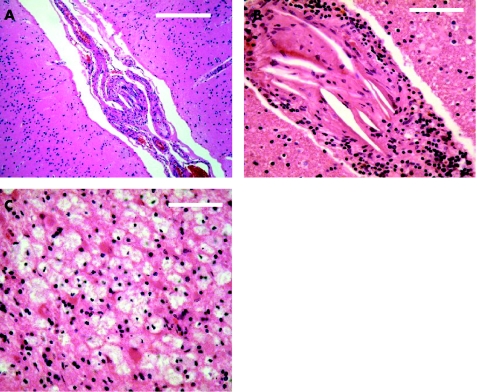
Figure 2 Brain histopathology. Small leptomeningeal (A) and perforating brain (B) arteries containing numerous cholesterol clefts and focal accumulation of lymphocytes in the adventitia. Lipid‐laden macrophages and reactive astrocytes in a small subcortical infarct (C). Scale bars: (A) 100 μm, (B) 50 μm, (C) 50 μm.Hematoxylin‐eosin stain.

An official website of the United States government
Here's how you know
Official websites use .gov
A
.gov website belongs to an official
government organization in the United States.
Secure .gov websites use HTTPS
A lock (
) or https:// means you've safely
connected to the .gov website. Share sensitive
information only on official, secure websites.
