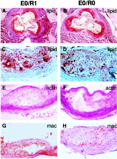
Figure 1.
Aortic root and arch lesions from 16-week-old Western diet-fed mice. (Left) apoE-deficient, Rag-1 heterozygote mouse lesions (E0/R1). (Right) apoE-deficient, Rag-1-deficient mouse lesions (E0/R0). (A and B) Oil red O staining for lipid in aortic root lesions (4× objective). (C and D) Oil red O staining for lipid in aortic root lesions (20× objective) reveals fibroproliferative lesions with fibrous caps and cholesterol clefts. (E and F) α-Actin antibody staining (dark magenta) in aortic arch fibroproliferative lesions demonstrates smooth muscle cells in the fibrous cap (20× objective). (G and H) Macrophage antibody staining (red-brown) in aortic arch lesions with necrotic cores (20× objective).

