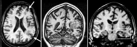
Figure 1 Middle panel: Coronal T1‐weighted image showing unilateral periventricular heterotopia. Note that the cortex above the lesion is abnormal, and probably contains areas of polymicrogyria. Left panel: Axial T1‐weighted image showing that the lesion involves mostly watershed areas at the frontal and occipital lobes (arrows). Right panel: Coronal T1‐weighted image showing atrophy of the left hippocampal formation (box).

An official website of the United States government
Here's how you know
Official websites use .gov
A
.gov website belongs to an official
government organization in the United States.
Secure .gov websites use HTTPS
A lock (
) or https:// means you've safely
connected to the .gov website. Share sensitive
information only on official, secure websites.
