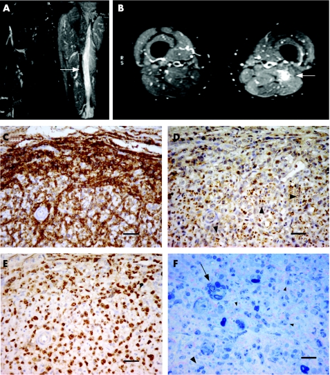
Figure 1 T2‐weighted magnetic resonance imaging of the thighs (A, B). High signal is present in the left sciatic nerve as compared with the right in coronal (A) and axial sections (B) (arrows), consistent with an infiltrating mass. High signal extends throughout the length of the sciatic nerve and is associated with swelling and oedema in the surrounding muscle. Fascicular biopsy of the sciatic nerve (C–F), immunostained with anti‐CD20 (C), PGP9.5 (D) and Ki‐67 (E). The endoneurium is heavily infiltrated with large pleomorphic cells immunoreactive for CD20 and some surrounding blood vessels (C). Remaining small, largely unmyelinated fibres, in Remak bundles (arrow heads), persist throughout the tumour (D). Ki‐67 staining shows a proliferation index of nearly 100% (E). 1‐μm resin sections show a few degenerating myelinated fibres (arrow) and a few persisting small myelinated fibres (large arrowhead) interspersed with the numerous abnormal tumour cells (small arrowheads) (F). Scale bars 40 μm (C–E) and 20 μm (F).

An official website of the United States government
Here's how you know
Official websites use .gov
A
.gov website belongs to an official
government organization in the United States.
Secure .gov websites use HTTPS
A lock (
) or https:// means you've safely
connected to the .gov website. Share sensitive
information only on official, secure websites.
