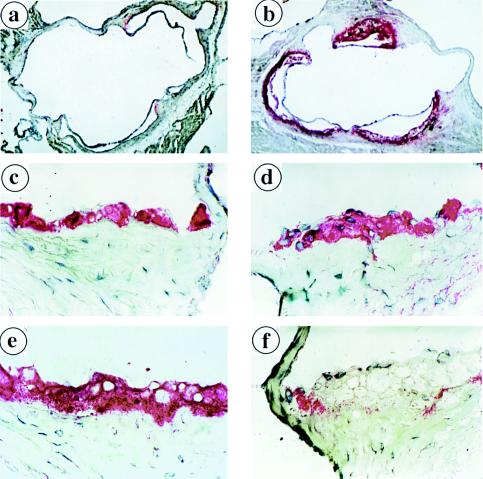
Figure 3.
Increased aortic atherosclerosis in C57BL/6 recipients of apoE(−/−) marrow. (a and b) Comparison of the extent of lipid staining by oil red O in representative sections of the proximal aorta of control apoE(+/+)→apoE(+/+) and experimental apoE(−/−)→apoE(+/+) mice after 13 weeks on the atherogenic diet (10×). Cryosections 10 μm thick were fixed with 4.0% paraformaldehyde, stained with oil red O, and counterstained with hematoxylin. (c and e) Serial sections from control animals. (d and f) Serial sections from apoE(−/−)→apoE(+/+) mice. (c and d) Immunocytochemical staining for macrophages reveals that foam cell lesions consist primarily of macrophages in experimental and control mice (63×). (e and f) Immunocytochemical staining of serial aortic sections for apoE (63×). ApoE staining in control mice colocalizes with macrophages, but the macrophages in the experimental mice do not stain with apoE.

