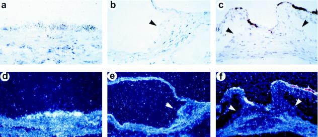
Figure 4.
Decreased apoE gene expression in the aortic root of apoE(−/−)→apoE(+/+) mice by in situ hybridization. The hybridization signals of the apoE antisense probe appears as black grains located in aortic foam cell lesion of apoE(+/+)→apoE(+/+) mice on bright field (a, 40×), or as white dots on dark field (d, 20×). (b and e) The sense probe did not produce hybridization with foam cell lesions under the same conditions on bright field (40×) or dark field (20×), respectively. (c and f) Absence of apoE expression in foam cell lesions of the proximal aorta in an apoE(−/−)→apoE(+/+) mouse after hybridization with the antisense probe on bright field (40×) and dark field (20×). The intense coloration of the borders of the aortic valve stumps visible in c and f is an artifact not related to hybridization-specific silver grain exposure.

