
Figure 5. Periodic loss of entrainment of pacemaker activity.
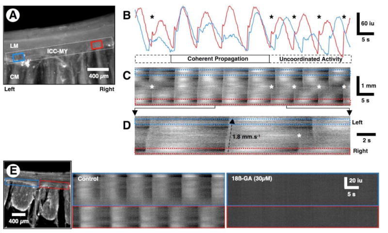
(A) Averaged Ca2+ fluorescence in a cross-sectional preparation. In this preparation, Ca2+ transients were measured only in ICC-MY. An ST map was constructed from the dotted white rectangle covering the ICC-MY network within the FOV. Ca2+ traces were also sampled in the ROIs (blue and red rectangles) near the opposite ends of the ICC-MY network. (B) Line traces taken from the red and blue ROIs in A. Note that pacemaker activity in ICC-MY is entrained and loses entrainment over a series of several cycles and is variable in amplitude with each cycle. (C) The ST map shows instances of propagated rhythmic activity along the entire length of the ICC-MY network (see also D). During the first two and last three cycles, pacemaker activity was not entrained (*) and two independent pacemaker regions emerged with similar frequencies. During the 3rd to 6th cycles, pacemaker activity propagated along the entire ICC-MY network in the FOV. (D) An expanded region of the ST map in C shows propagation across the ICC-MY network during the first two cycles (bottom to top in the ST map) and loss of entrainment of activity during the 3rd cycle (*). (E) ST maps were constructed from each of the blue and red rectangles along the ICC-MY network of the preparation shown on the left. Pacemaker activity in the two regions of the ICC-MY network occurred at similar frequencies, but the activity was out of phase. 18ß-glycyrrhetinic acid (18ß-GA, 30 μM) blocked all activity in the ICC-MY network.