
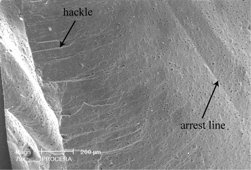
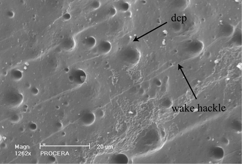
Fig. 5.
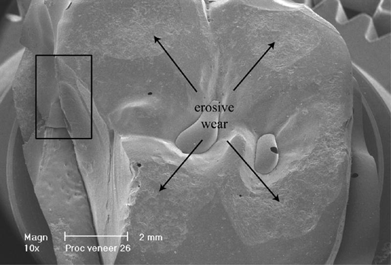
a: Occlusal view (at 10x) of the Procera AllCeram crown showing the veneer failure on the palatal-mesial cusp. Major wear is visible on all four cusps. The frame near the mesial gingival margin is magnified in Fig.5b and Fig.5c
b and c: Higher magnifications within the frame (Fig.5a) near the interproximal mesial margin show hackle and wake hackle indicating the direction of crack propagation towards the margin.