
Figure 1. Cell binding, or lack of binding, to lectin derivatized bead(s).
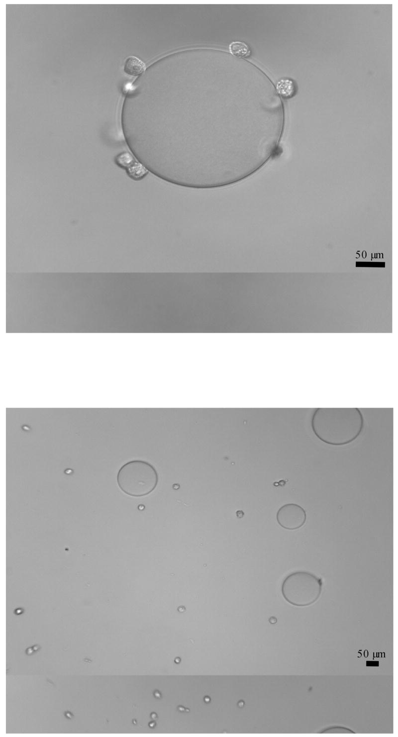
Top: Example of positive cell-bead binding. Fixed CCL-220 cells (small spheres) binding to a single DBA-derivatized agarose bead. Original magnification 400x.
Bottom: Example of negative cell-bead binding. Fixed CCL-220 cells (small spheres) not binding to DBA-derivatized agarose beads (large spheres) in the presence of 0.75M N-acetyl-D-galactosamine. Original magnification 200x.