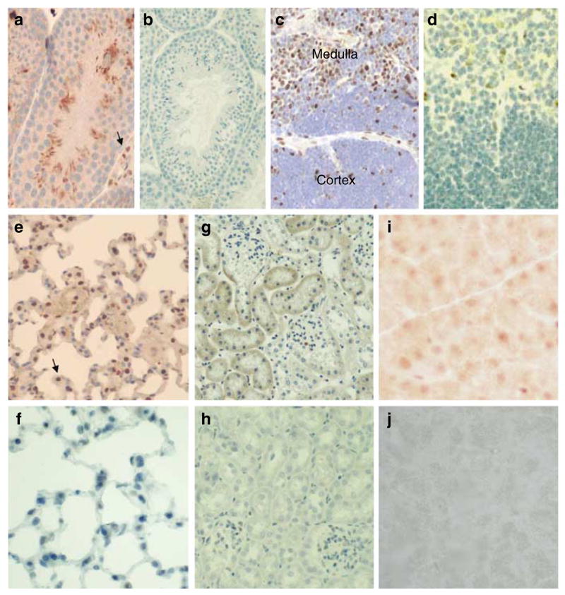
Figure 2.

Immunohistochemical detection of Dmp1 in tissues. (a) Immunohistochemical analysis of Dmp1 localization visualized with DAB (brown staining) in the testis and counterstained with hematoxylin. (b) Testis from a Dmp1−/− mouse stained with RAX antibody followed by hematoxylin counterstain. (c) Thymus with positive Dmp1 staining in the medulla. (d) Thymus from a Dmp1−/− mouse stained with RAX, then with hematoxylin counterstain. (e) Dmp1 staining of the lung. Dmp1 localization is visualized with DAB in alveolar macrophages and epithelial cells. (f) Lung from a Dmp1−/− mouse stained with RAX, then with hematoxylin counterstain. (g) Dmp1 staining in the kidney. (h) Kidney from a Dmp1−/− mouse. (i) Weak Dmp1 staining in the pancreas. (j) Pancreas from a Dmp1−/− mouse.
