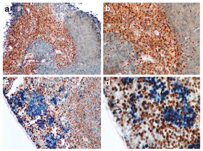
Figure 3.

Immunohistochemical detection of Dmp1 in normal thymus and spleen. (a, b) Thymic tissue sections were stained with the Dmp1 and Ki67 antibodies, using peroxidase-conjugate and DAB for Dmp1 (brown) and alkaline phosphatase-conjugate for Ki67 (blue). Note that the staining for Dmp1 and Ki67 are very exclusive. (a) × 10; (b) × 20. (c, d) splenic sections from formalin-fixed and paraffin-embedded mouse tissue were stained with the Dmp1 and Ki67 antibodies, using peroxidase-conjugate and DAB for Dmp1 (brown) and alkaline phosphatase-conjugate for Ki67 (blue). Dmp1 nuclear expression is found mainly in the germinal centers of the organ, while Ki67 was expressed in mononuclear cells in the subcortical area. Magnification, (c) × 10; (d) × 20.
