
Figure 4.
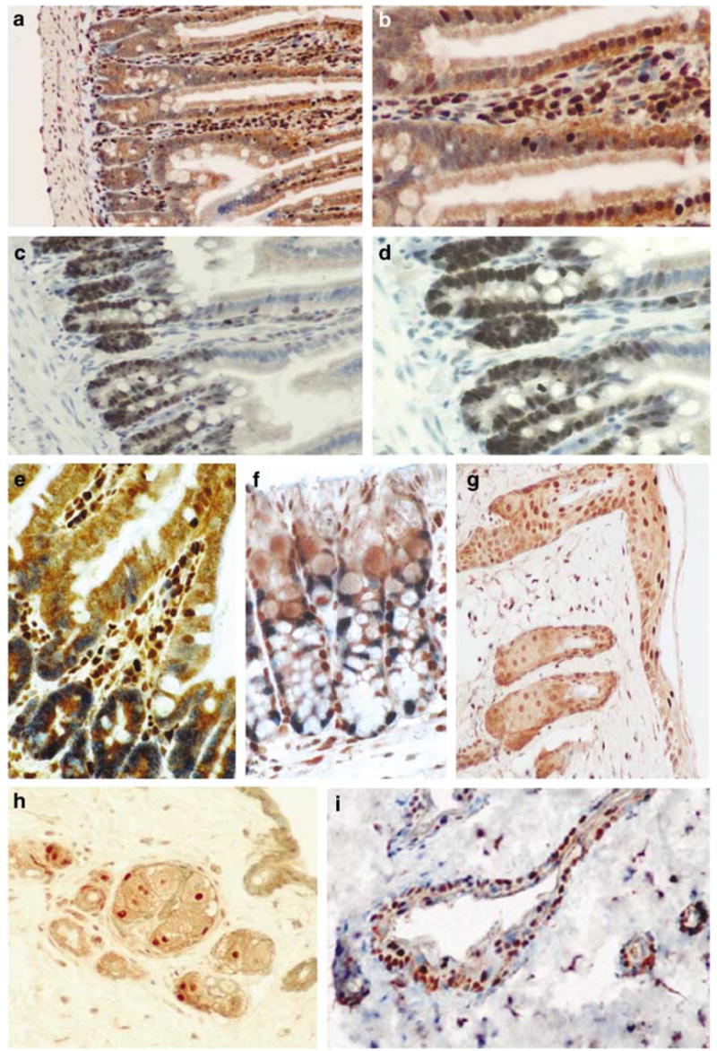
Mutually exclusive staining of Dmp1 and Ki67 in normal intestines (a–f) and skin (g–i). (a, b) Staining with the Dmp1 antibody in the small intestine of a wild-type mouse using peroxidase-conjugate and DAB substrate (brown). Magnification: (a) ×10; (b) × 20. (c, d) Showing the positive expression of Ki67 in the small intestine (dark blue) (c, ×10; d, ×20). (e) Showing double staining with Dmp1 and Ki67 in small intestine (× 40). (f) Showing Dmp1 (brown) and Ki67 (blue) double staining in mouse colon (× 40). (g) Showing Dmp1 staining in the skin (brown, × 20). (h) Showing Ki67 staining of the skin in a cross section of a hair root (brown, ×20). (i) Showing the Dmp1 (brown) and Ki67 (blue) double staining of the skin (× 20). The Dmp1 expression is more localized at the epidermis.