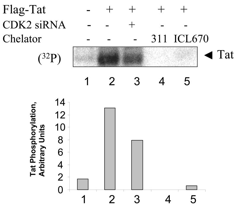
Figure 6. Iron chelators reduce HIV-1 Tat phosphorylated in cultured cells.
HeLa cells were infected with recombinant adenovirus expressing Flag-tagged Tat as described in Methods (lanes 2–5). Lane 1, control uninfected cells. HeLa cells were transfected with siRNAs targeting CDK2 (lane 3) or treated with 10 μM 311 or 100 μM ICL670. At 48 hours post infection cells were labeled with (32P)-orthophosphate for 2 hours with the addition of 1 μM okadaic acid. Whole cell extracts were prepared and Tat was immunoprecipitated with anti-Flag monoclonal antibodies, resolved on 15% Tris-Tricine SDS-PAGE and detected by Phosphor Imager. Phosphor Imager quantification is shown in the lower panel.

