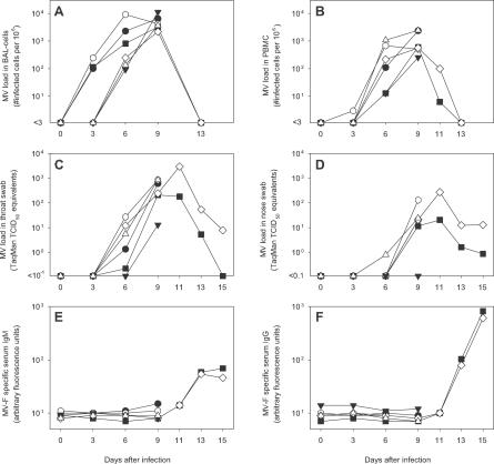
Figure 1. MV Replication and Specific Antibody Responses in Macaques at Different Time Points after Infection.
Virus isolation from BAL cells (A) and PBMCs (B); virus detection by TaqMan RT-PCR in throat (C) and nose (D) swabs; MV fusion protein (F)–specific serum IgM (E) and IgG (F) responses as determined by FACS-measured immunofluorescence. Symbols indicate rhesus macaques #R1 (•), #R2 (▪), #R3 (▾), and cynomolgus macaques #C1 (○), #C2 (⋄), and #C3 (Δ).

