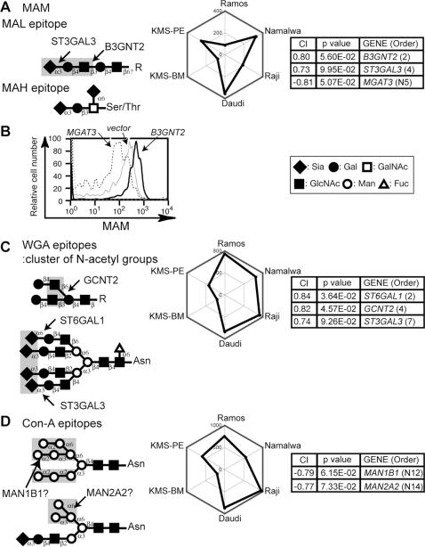
Figure 5. CIRES analyses of staining profiles obtained using lectins that recognize multiple glycan structures and have unknown epitope expression-regulating enzymes.
Presentation is the same as in Fig. 2 except that the plant lectins used were (A–B) MAM, (C) WGA, and (D) Con-A. The epitopes of the two different lectins of MAM, MAL and MAH, are illustrated separately. WGA essentially recognizes a cluster of N-acetyl groups, as indicated, and thus required Neu5Ac as a Sia species. Con-A recognizes mannose-containing glycans with varying affinities. High-mannose-type glycans (upper diagram in (D)) bind best to this lectin. (B) Namalwa cells were infected with retroviruses encoding various GlcNAc transferases. The MAM staining patterns of the EGFP-positive populations of each infectant are shown. MGAT5 overexpression did not shift the staining pattern in comparison with the vector control (data not shown).

