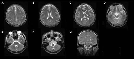
Figure 1 An example of magnetic resonance images obtained from a patient with a left subcortical infarct on the first week after the onset of stroke. From axial T2 weighted images (A–F) and the coronal fluid attenuated inversion recovery image (G), the subcortical infarct involving the posterior limb of the internal capsule is observed (white arrows). Beyond the primary lesion, there is no hyperintense signal.

An official website of the United States government
Here's how you know
Official websites use .gov
A
.gov website belongs to an official
government organization in the United States.
Secure .gov websites use HTTPS
A lock (
) or https:// means you've safely
connected to the .gov website. Share sensitive
information only on official, secure websites.
