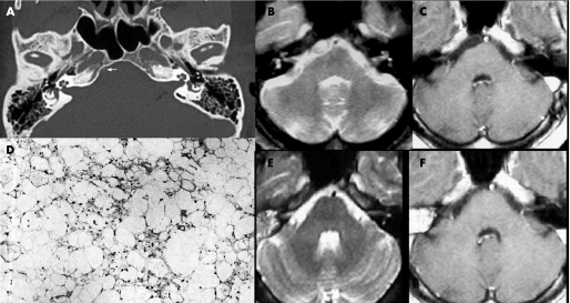
Figure 1 (A) CT (bone window) shows a subtle stalk‐like structure projecting from the clivus (arrow). (B, C) Axial MRI ((A) T2 weighted image (T2WI); (B) post‐contrast T1 weighted image (T1WI)) revealed a small well circumscribed mass in the prepontine region, mildly compressing the pons. The lesion appears hyperintense on T2WI (A) and hypointense on T1WI without contrast enhancement (B). (E, F) One year follow‐up axial MRI demonstrates no recurrence of the mass on T2WI (A) and on post‐contrast T1WI (B). (D) Light photomicrograph: hypocellularity of the physaliphorous cells with a lobular growth pattern, eosinophilic and vacuolated cytoplasm with a mixomatous matrix, absence of mitoses and of cellular pleomorphism (haematoxylin‐eosin ×200).

An official website of the United States government
Here's how you know
Official websites use .gov
A
.gov website belongs to an official
government organization in the United States.
Secure .gov websites use HTTPS
A lock (
) or https:// means you've safely
connected to the .gov website. Share sensitive
information only on official, secure websites.
