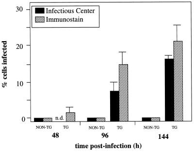
Figure 4.
Detection of MV in primary neuron cultures. Primary neurons cultured from transgenic (TG) (line 52) and nontransgenic (NON-TG) embryonic hippocampi were infected with MV–Edmonston at a multiplicity of infection = 3, 48 hr after culturing. At various times after infection, neurons were harvested for infectious center analysis or immunostained with SSPE serum. Supernatants were also collected for plaque assay. For the immunostaining, 10 fields per time point were counted, and standard deviations are shown.

