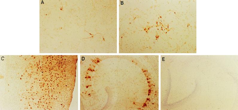
Figure 5.
Spread of MV in primary neurons and in vivo. (A) CD46+ primary neurons, infected for 48 hr with MV–Edmonston. (B) CD46+ primary neurons 96 hr after infection. Coverslips were fixed with 1:1 acetone:methanol and immunostained with a human SSPE serum as described. (C–E) NSE–CD46 neonatal (<24 postnatal) transgenic and nontransgenic mice were inoculated intracranially with 105 pfu of MV–Edmonston and brain sections were immunostained for MV as described. Immunohistochemical staining for MV antigens in the cortical region (C) and in the hippocampus (D) of a representative, NSE–CD46 transgenic mouse infected with MV 10 days previously (×100). (E) Hippocampus of a nontransgenic mouse, inoculated 10 days previously (×100).

