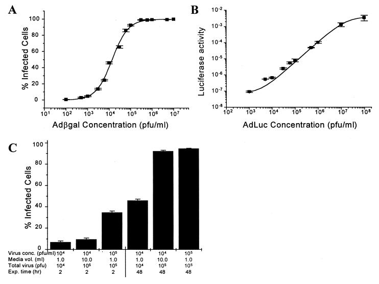
Figure 1.
Influence of virus concentration and exposure time on recombinant adenovirus infection in primary cultures of adult rabbit ventricular myocytes at 37°C. (A) Percentage of cells positive for β-gal after 48 hr exposure to Adβgal (1.0 × 10⁵ cells per culture dish). Control, uninfected cells did not stain positive for β-gal activity. (B) Normalized luciferase activity (mg/mg protein) after 48 hr exposure to varying concentrations of AdLuc (2.0 × 10⁵ cells per culture dish). Luciferase activity in control, uninfected cells was 1.2 × 10⁻⁹ mg/mg protein. Solid lines are logistical regression curves drawn through data. (C) Percentage of Adβgal-infected cells after 2 hr or 48 hr virus exposure (1.0 × 10⁵ cells per culture dish). Columns 1–2 and 4–5 are the same virus concentration, and columns 2–3 and 5–6 are the same total virus amount. (n = 3.)

