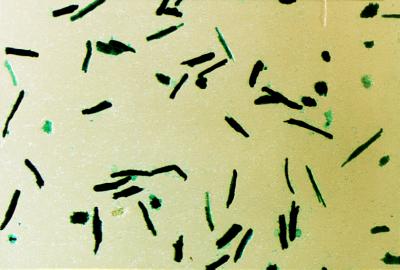
Figure 4.
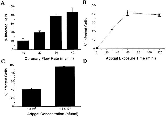
Adβgal delivery to intact, Langendorff-perfused rabbit hearts at 37°C. (A) Percentage of myocytes positive for β-gal following 2.0 hr infections with 1.0 × 10⁸ pfu/ml Adβgal at coronary flow rates ranging from 10 to 40 ml/min. (B) Effect of virus perfusion time on infection with 1.0 × 10⁸ pfu/ml Adβgal at 30 ml/min. The exposure time varied from 1.67 min to 2.0 hr with the virus-free rinse duration adjusted to maintain a total Langendorff perfusion time of 180 min. (C) Effect of virus concentration on infection rates with 60 min Adβgal perfusion at 30 ml/min. (n = 3.) (D) 5-Bromo-4-chloro-3-indolyl β-d-galactopyranoside (X-gal)-stained myocytes from a heart infected with 1.6 × 10⁹ pfu/ml of Adβgal during Langendorff perfusion.


