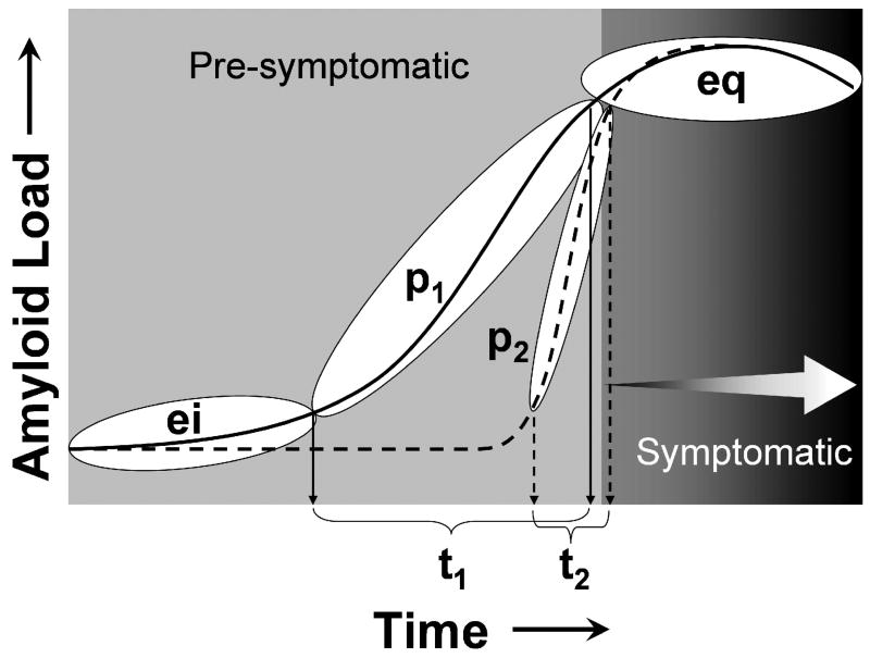
Figure 4.
Schematic of the hypothetical progression of Aβ deposition over time from the early initiation (ei) phase, to the continuously progressive (p) phase, and finally the late equilibrium (eq) phase. Subjects may experience either a long (p1/t1) or brief (p2/t2) progressive phase of Aβ deposition, which snapshots such as shown in Figure 3 can capture at a single point in time. Cognitive symptoms may not be evident until the equilibrium (eq) phase (MCI+, MCI++, and AD in Figure 3), but the cascade of pathological events that may lead to these symptoms (i.e. neurofibrillary pathology, synapse and neuron loss) may be initiated during the progressive phase (p) (figure from [94])

