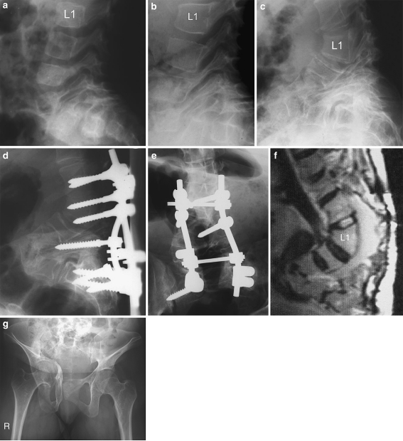
Fig. 1.
a Radiograph of the lumbar spine of patient 1 at the age of 3 years and 5 month shows a lumbar hyperlordosis of 92° (from L3 to S1) and spondylolisthesis due to elongation of lumbar pedicles, markedly of L3, L4, and L5. b At 7 years and 11 month of age, lumbar pedicle elongation and consecutive spondylolisthesis show a rapid progression. c Radiograph of patient 1 at 17 years of age shows angular hyperlordosis of 126° (from L3 to S1) and spondylolisthesis as a result of extreme elongation of lumbar pedicles. d, e Radiographs of patient 1 after laminectomy and postero-lateral fusion using Cotrel–Dubousset instrumentation. f A recent MRI scan of the lumbar spine of patient 1 at the age of 28 years shows no significant progression of the lumbar deformation. g A current antero-posterior radiograph of the pelvis of patient 1 shows the pelvic deformity with acetabular protrusion and fracture of the right acetabulum

