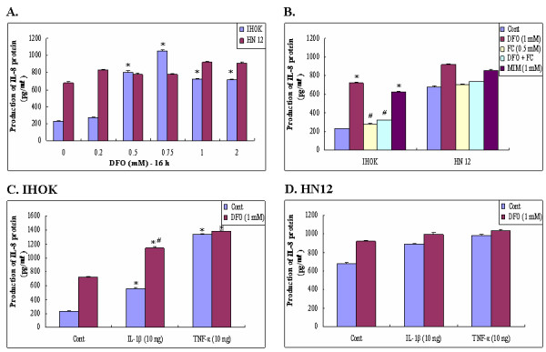
Figure 1.

Effects of Iron chelator on IL-8 production in immortalized (IHOK) and malignant human oral keratinocytes (HN12). Cells were treated for 16 h with the indicated concentrations of DFO (0.2–2 mM) in IHOK and HN12 cells (A), or DFO (1.0 mM), FC (0.5 mM), MIM (1.0 mM) in IHOK and HN12 cells (B), IL-1 β (10 ng/ml), TNF-α (10 ng/ml), or combinations thereof in IHOK (C) and HN12 (D) cells. Levels of IL-8 secretion were determined by ELISA. Results are expressed as means ± SD of three independent experiments. Numbers below the gels represent the intensity of IL-8 mRNA relative to GAPDH mRNA.*: Statistically significant difference compared to control group, p < 0.05. #: Statistically significant difference compared to DFO group, p < 0.05.
