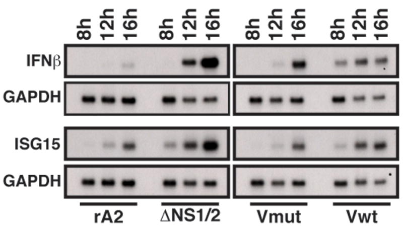
Figure 3.

Accumulation of IFNβ and ISG15 mRNA in rRSV-infected cells. A549 cells were infected with the indicated viruses at a MOI of 3. At 8, 12, and 16 h p.i., total RNA was isolated from the infected cells and subjected to Northern blot analysis using radiolabled DNA probes to IFNβ (top) or ISG15 (bottom). Bands were visualized by autoradiography. After exposure, the blots were stripped and reprobed with a radiolabeled probe for GAPDH as a loading control (lower panels).
