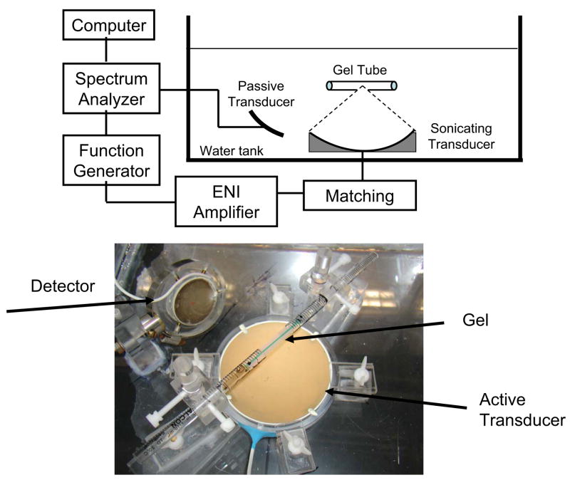
Fig 3.
(a) Side-view schematic diagram of experimental setup for the measurement of the collapse threshold of microbubbles in gel tunnels. Microbubbles were injected into the tunnel before the insonation. Two transducers were used and placed in the water tank: one generates the ultrasound waves and the other one listens to the acoustic emission. (b) Picture of the tray with the gel tube inserted between the two polystyrene tube segments.

