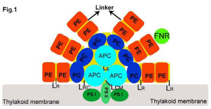
Fig 1.
Structural model of a tricylindrical hemidiscoidal phycobilisome (2, 3). The three sky blue circles represent the tricylindrical core APC, and two bottom cylinders attach to the thylakoid membrane (grey rectangle) with LCM. Six rods are arranged by PC (blue circle), and PE (red circle), and attached FNR (grass green circle) with LR from inner to outer part. LRC is the linker between core and rod. All linkers are represented by yellow discs located in each rod.

