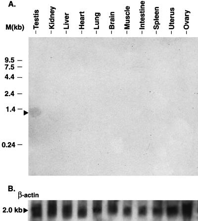
Figure 2.
(A) Northern blot of various tissues mRNAs probed with FA-1 cDNA. Two micrograms of poly(A)+ RNA isolated from each tissue was separated on a 1.2% denaturing agarose/formaldehyde gel and transferred to nitrocellulose membrane. The membrane was prehybridized with QuickHyb solution, incubated (56°C, 2 hr) with 32P-labeled FA-1 cDNA probe, washed, and exposed to x-ray film for 24 hr to 3 weeks. (B) After the membrane was stripped of the FA-1 probe, it was rehybridized (65°C, 2 hr) with 32P-labeled β-actin probe, washed, and exposed as above.

