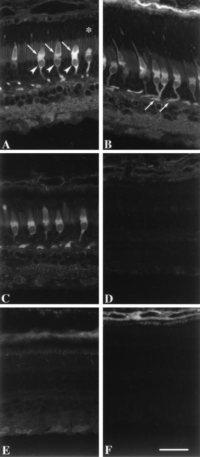
Figure 3.
Immunofluorescence localization of GCAP2 in monkey retina. (A) GCAP2 immunolabeling with UW50 is strongest in the cone inner segments (arrows), somata (arrowheads), and synaptic terminals. Cone nuclei are negative images. Weak immunolabeling is present in the rod and cone outer segments (denoted by asterisk), rod inner segments, and neurons in the inner retina. (B) Although individual cones are rarely visible in their entirety within a single image plane, their cellular processes can be visualized with a projection of a z series. Arrows denote cone synaptic terminals. (C) Addition of purified GCAP1 (25 μg/ml) to anti-GCAP2 polyclonal antibodies produces minimal decrease in GCAP2 immunoreactivity, verifying that this antibody does not cross-react with GCAP1. (D) Addition of GCAP2–7 (25 μg/ml) to anti-GCAP2 polyclonal antibodies abolishes GCAP2 immunoreactivity. Sections incubated in preimmune serum (E) or buffer without anti-GCAP2 (F) show no immunolabeling of cones or other retinal cell types. The choroid (at the top) is weakly reactive with the secondary antibody. (Bar = 20 μm.)

