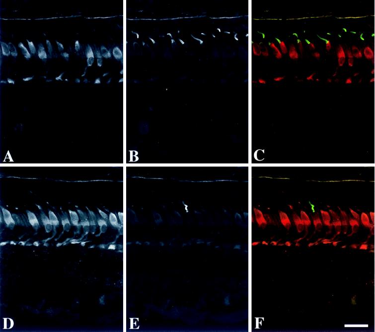
Figure 4.
Immunofluorescence localization of GCAP2 and cone opsins in monkey retina. (A–C) Localization of GCAP2 and red/green cone opsin. (A) The cones are immunolabeled with anti-GCAP2 (UW50), with the strongest labeling in the inner segments, somata, and synaptic terminals. Some inner retinal neurons are weakly labeled. (B) Anti-red/green cone opsin labels the majority of cone outer segments. (C) Double labeling with anti-red/green cone opsin (green) and anti-GCAP2 (red) demonstrates that the red/green cones are immunopositive for GCAP2. (D–F) Localization of GCAP2 and blue cone opsin. (D) The cone photoreceptors are immunolabeled with anti-GCAP2. (E) Anti-blue cone opsin labels a single cone outer segment. (F) Double labeling with anti-blue cone opsin (green) and anti-GCAP2 (red) demonstrates that the blue cone is immunopositive for GCAP2. (Bar = 20 μm.)

