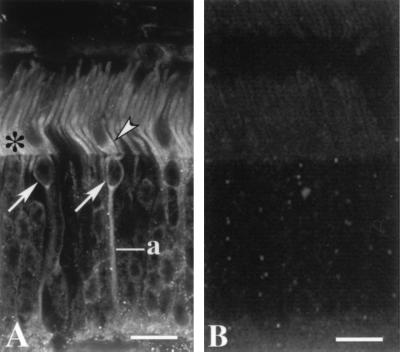
Figure 5.
Immunofluorescence localization of GCAP2 in bovine retina. (A) Both rod and cone photoreceptors are labeled. In rods, labeling is strongest in the inner segments (asterisk). The cone inner segments are substantially wider than rod inner segments and the cell bodies of cones are restricted to the outermost tier of the outer nuclear layer. Labeling is present in the myoid region of a cone inner segment (arrowhead), cone somata (arrows) and a cone axon (a). Labeled photoreceptor synapses are at the bottom. (Bar = 15 μm.) (B) Addition of GCAP2–7 (25 μg/ml) to anti-GCAP2 polyclonal antibodies abolishes GCAP2 immunoreactivity.

