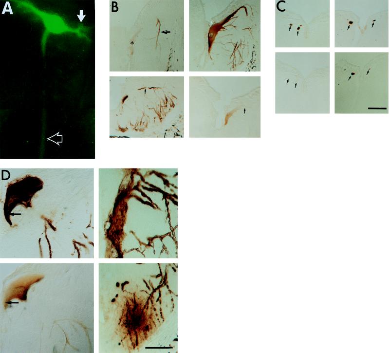
Figure 3.
Distribution and phosphorylation of full-length htau in lightly versus heavily expressing ABCs. (A) GFP fluorescence of a single ABC in the living lamprey hindbrain. GFP–htau is distributed throughout the dendrites (solid arrow), soma, and axon (open arrow). (B) Adjacent transverse sections through the somata and dendrites of lightly (Upper, 17 days p.i.) and heavily (Lower, 8 days p.i.) expressing ABCs in the hindbrain, showing phosphorylation of dendritic htau at the PHF-1 and TAU-1 sites. Both cells expressed three-repeat full-length htau without GFP. Sections were stained with PHF-1 (Left) and TAU-1 (Right). (Upper Right) Section was pretreated with AP to show total htau distribution. Note that normal morphology and an even somatodendritic distribution of total htau is combined with the selective accumulation of PHF-1-positive htau in the distal dendrites of a lightly expressing ABC (Upper, arrow) relative to the soma (Upper, asterisk). A heavily expressing ABC (Lower Left) shows PHF-1 staining throughout the soma and dendrites (arrow). Staining for TAU-1 is absent from dendrites (Lower Right, arrow). (C) Transverse sections through ABC axons ≈1 mm from their parent somata in the caudal hindbrain. (Left) Adjacent sections through the axons of three ABCs 8 days p.i. expressing three-repeat full-length htau without GFP; (Right) Adjacent sections from another lamprey examined 11 days p.i. expressing the same construct in which all of the axons (arrows) from ABCs were judged to be staining heavily. Note the absence of PHF-1 staining in the 8-day axons (Lower Left, arrows) and its strong presence in 11-day axons (Lower Right). (D) In heavily staining ABCs examined 9 days or later p.i., staining with both PHF-1 (Upper Left) and TAU-1 (Lower Left) was observed throughout the cells. Most htau accumulations were distributed in a clumped, uneven pattern in the soma and were hyperphosphorylated (i.e., PHF-1-positive and TAU-1-negative; Left, arrows). Some of these ABCs were undergoing degeneration (Upper Right); these often exhibited distal dendrites surrounded by extracellular htau accumulations in the adjacent neurophil (Lower Right, ∗). (Bars = 50 μm.)

