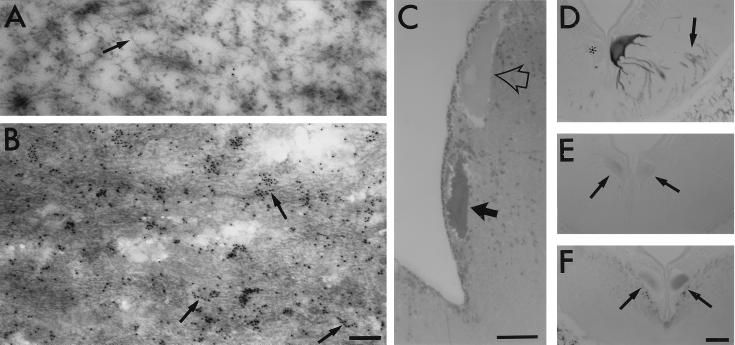
Figure 4.
Incorporation of htau into 10- to 15-nm filaments in ABCs. (A–C) ABCs expressing GFP-tagged htau were identified by the localization of fluorescent cells in freshly isolated brains before fixation. htau-expressing ABCs targeted in this manner stained more strongly with toluidine blue (C, solid arrow) than did adjacent nonexpressing cells (open arrow). Ultrastructural analysis of adjacent thin (80 nm) sections through these cells showed that htau-expressing ABCs contained densely packed bundles of 10- to 15-nm filaments (B), which are absent from the somata and dendrites of nonexpressing cells in the same section (A). Many of the filaments in the htau-expressing cells became decorated with 10-nm colloidal gold particles (B, arrows) when thin sections embedded in LR White were probed with an antiserum directed against GFP (CLONTECH) followed by a gold-labeled secondary antibody (Sigma). The patchy labeling in B is probably due to poor penetration of immunogold and the preferential decoration of filaments on the section surface. Note that NFs in nonexpressing cells were not labeled at all (A, arrow). [Bars = 200 nm (A and B) and 50 μm (C).] (D–F) Adjacent sections through the somata and dendrites of a pair of ABCs, one of which expresses htau (darkly stained cell at right) and one that does not (∗), were stained with the monoclonal antibodies PHF-1 (D), RMO44 (which selectively recognizes non phosphorylated NFs; E), and RMO62 (which recognizes a phosphorylated epitope on lamprey NF protein; F). Note that no increase in staining for somatodendritic NFs is visible in expressing cells (E and F, arrows) whose dendrites stain strongly for PHF-1 (D, arrows). (Bar = 50 μm.)

