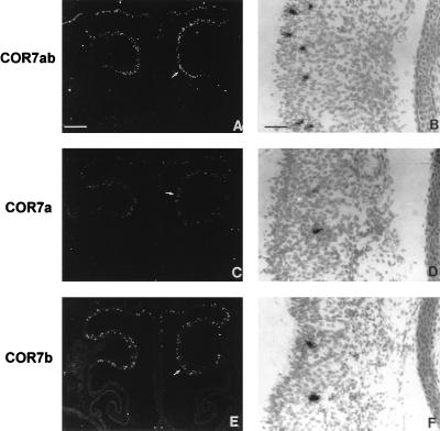
Figure 3.
mRNA localization of COR7a and COR7b in the olfactory epithelium. Serial adjacent sections (14 μm) of the nasal cavity at E16 were hybridized with 35S-labeled antisense RNA probes prepared from COR7a (C and D), COR7b (E and F), and COR7ab (A and B) genes. B, D, and F represent enlargements of the olfactory epithelium covering the olfactory conchae (arrows in A, C, and E). With dark-field illumination, positive cells appear as white spots on a black background. At bright-field and higher magnifications, positive cells are covered with dense silver grains. Note the random distributions of COR7a and COR7b transcripts within the olfactory epithelium and their localization in the middle layer of the epithelium that correspond to the nuclei of olfactory neurons. The sum of labeled cells with COR7a (≈110 cells) and COR7b (≈135 cells) corresponds approximately to the number of positive cells detected with the nonspecific probe COR7ab (≈255 cells), indicating that there is no coexpression of the two genes in the same cell. [Bars = 270 μm (A, C, and E) and 8 μm (B, D, and F).]

