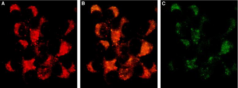
Figure 3.
MIF is colocalized in the insulin-containing granule of β cells. INS-1 cells were stained with anti-insulin (A), anti-insulin and anti-MIF (B), or anti-MIF IgG (C) and revealed by immunofluorescence (Texas Red for the anti-insulin antibodies and fluorescein isothiocyanate for the anti-MIF). Insulin granules are red, MIF staining is green, and the orange-yellow staining corresponds to granules containing MIF and insulin. Fluorescence images were obtained with confocal laser scan microscope. (×60.)

