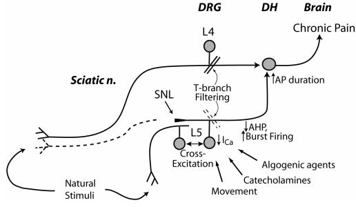
Figure 12.
Schematic diagram depicting the contribution of reduced sensory neuron Ca2+ current (ICa) to neuropathic pain. Injured nerve tissue distal to the spinal nerve ligation (SNL) undergoes Wallerian degeneration (dotted line), while neurons from the fifth lumbar (L5) dorsal root ganglion (DRG) are activated by movement, catecholamines, other algogenic agents and cross-excitation from adjacent intact neurons. Reduced ICa shortens afterhyperpolarizations (AHPs), which contributes to burst firing. Loss of ICa impairs natural signal filtering at the T-branch, where the stem axon splits into spinal nerve and dorsal root branches. Reduced ICa also prolongs action potential (AP) duration, which may increase excitatory synaptic transmission in the dorsal horn (DH). Spared nerves transmit activity evoked by stimulation in the receptive field, and these signals encounter DH neurons that are sensitized by L5 input. From McCallum et al (48), with permission.

