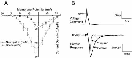
Figure 4.
Inward low-voltage activated Ca2+ currents measured by patch-clamp recording in dissociated sensory neurons decrease after chronic constriction injury. (A) Current-voltage plot of average data for medium sized-neurons shows a loss of peak current in injured neurons from animals with neuropathic pain, and particularly a loss of current at low voltages. (B) Presentation of a voltage command in the from of an action potential (top) produces current through low-voltage activated Ca2+ channels (bottom) that is substantially reduced in an injured neuron compared with a control neuron. From McCallum et al (49), with permission.

