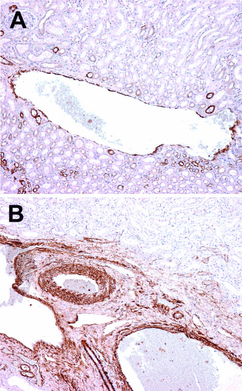
Figure 2.
Positivity for smooth muscle actin in control kidney. A, Large interlobular vein with muscle layer of varying thickness. B, Arcuate vein with well-developed muscle wall. The wall of the corresponding artery is thicker, with densely packed muscle cells. Streptavidin-biotin peroxidase; original magnification ×100.

