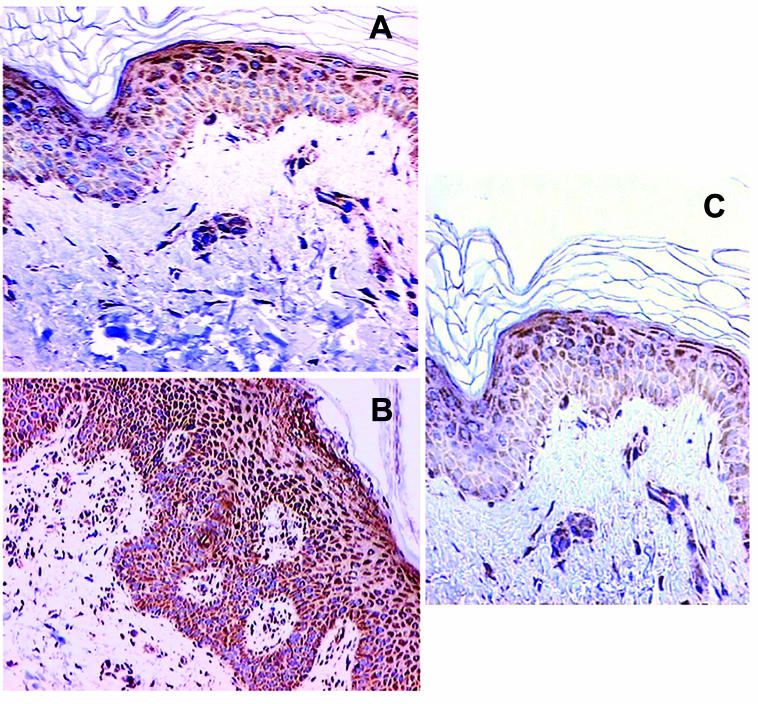
Figure 3.
Immunohistochemical staining for Bak. (A) Intense Bak staining mainly in suprabasal layers of normal skin with granular layer being stained slightly stronger than the spinous and basal layers ( × 400). (B) Strong diffuse Bak staining in involved psoriatic skin with granular layer being stained slightly stronger than the spinous and basal layers ( × 200). (C) Intense Bak staining predominately in the suprabasal layers of uninvolved psoriatic skin, with accentuation in upper layers ( × 400).

