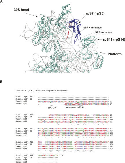
FIGURE 1.
Structure, ribosomal location, and sequence alignments of ribosomal protein S7 (rpS5). (A) Location of rpS7 within the head of the E. coli 30S ribosomal subunit. PDB file 1VS7 was used for modeling. Image was produced using Deep View 3.7 software. The rpS7 protein is in blue, and its 20 amino-terminal amino acids are in red. (B) Sequence alignments of E. coli strain K12 rpS7 (GenBank accession no. NP_417800), E. coli strain O6:H1/CFT073 (NP_755978), S. cerevisiae rpS5 (NP_012657), and H. sapiens rpS5 (NP_001000). The pI value of the N-terminal extension of yeast rpS5 is shown, and the peptide used to elicit anti-human rpS5 antibodies is underlined.

