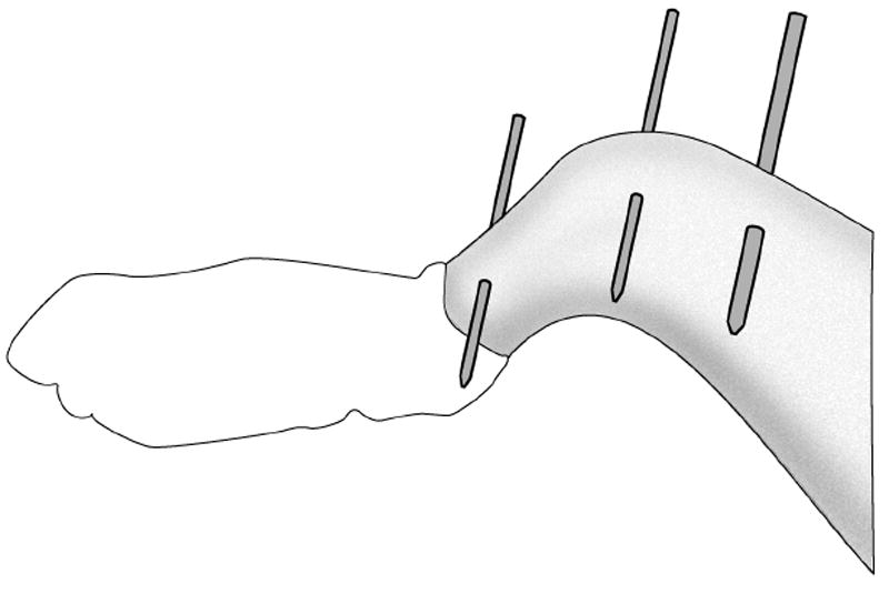
Figure 1.

Placement of two tibia pins and one femur pin in rabbit leg. The pins are subsequently cut to about 10mm on each side of the limb. These pins are used to hold and reproducibly position the leg in the knee testing device for tests at multiple time points.
