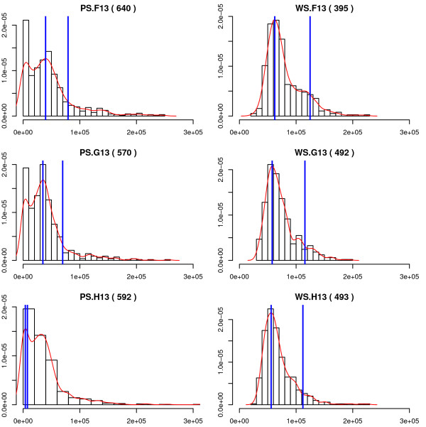
Figure 4.
DNA content of poorly-segmented (PS) and well-segmented (WS) objects. Each plot is a histogram of total DNA content from a single representative DMSO vehicle-treated well (F13, G13, or H13). The left column shows DNA content of poorly-segmented objects; well-segmented objects from the same wells are shown in the right column. The number in parentheses above each plot indicates the total number of objects included in the histogram. The red curve is a smoothed fit to the observed distribution. The blue lines were placed at the mode of the fitted distribution (the presumptive G0/G1 peak), and at twice the mode (the expected location of the G2/M peak). Note the poor estimates of the location of the G0/G1 peak in the poorly-segmented-class histograms, due to the large debris peak at small DNA content.

