
Figure 5. Head vasculature in double-null embryos at e9.5 and e10.5.
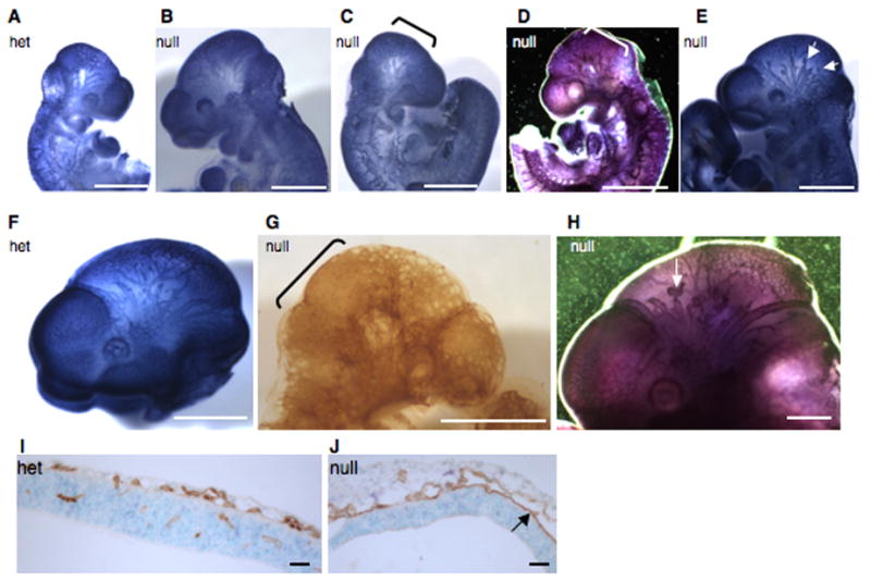
A–E. Pecam-1 staining of e9.5 heads. Note a regular pattern of small and large blood vessels in a heterozygous (A) and in a grossly normal double-null embryo (B), compared with a syncytial appearance of small head vessels in a morphologically defective (C) and a grossly normal (D) double-null embryo. Note blunt-ended and interrupted vessels in another grossly normal double-null embryo (E).
F–H. Pecam-1 staining of e10.5 heads. Note a regular pattern of small and large vessels in a heterozygous (F) embryo as opposed to syncytial appearance of small vessels and the scarcity of large vessels in a morphologically abnormal double-null embryo (G), as well as short, blunt-ended and interrupted head vessels in a grossly normal double-null (H). Brackets highlight syncytial endothelial sheets in C, D and G and arrows point to blunt-ended vessels in E and H. Scale bars are 1 mm in all except G, where scale bar is 240 μm.
I–J. Transverse sections through heads of a heterozygous (I) and a double-null (J) embryo at e10.5 show the presence of small vessels in the head and vessel invasion into the neural fold in the heterozygote (I). In the double-null (J) the vessels appear dilated, resulting from two sheets of endothelial cells touching each other in some places. Also note the lack of vascular invasion into the neural fold in the double-null (J). Scale bars are 65 μm.