
Figure 6. Extraembryonic vascular defects in EIIIA/EIIIB-double-null embryos.
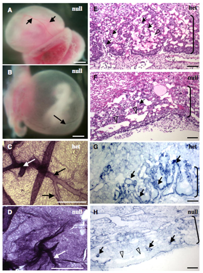
A–D. Yolk sacs. A, B. EIIIA/EIIIB-double-null yolk sacs showing normal (A) and defective (B) vasculature. Yolk sac in A has large (arrows) and small blood vessels while the one in B has only one large vessel (arrow) and abnormally patterned small vessels. C–D. A heterozygous and a double-null yolk sac stained with antibody to Pecam-1. White arrows point to vitelline vessels and black arrows point to large yolk sac blood vessels. Notice the size and the regular pattern of yolk sac vessels in C, and syncytial appearance of vasculature and absence of large blood vessels in D. Scale bars are 1 mm.
E–H. Placental labyrinths (brackets) in heterozygous (E and G) and double-null (F and H) embryos. Note extensive embryonic vasculature permeating the labyrinthine layer in E and the scarcity of embryonic blood vessels in F. Filled arrows point to embryonic and open arrowheads to maternal vessels.
G, H. Vascular pattern in the labyrinth layer is revealed with an antibody to laminin1. Note abundant finger-like vascular projections in heterozygous (G) but not double-null (H) placenta. Scale bars are 65 μm.