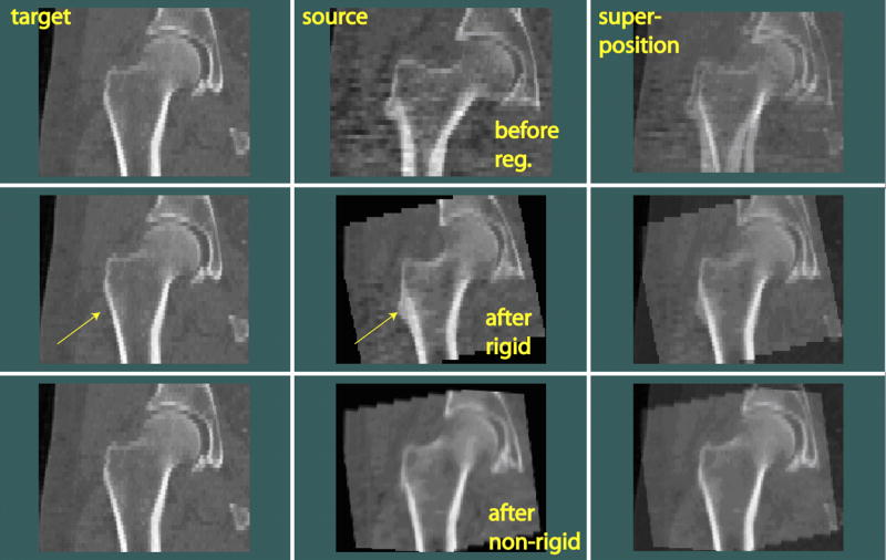
Fig. 4.

Rigid and non-rigid inter-subject registration of hip images (in coronal view). The source scan was rigidly transformed and warped toward the target scan. Arrows indicate the location where anatomical differences between the target and source can be easily seen.
