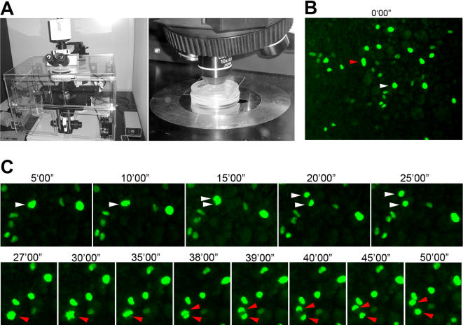
Figure 3. Time-lapse imaging of cell divisions in the ventral mesoderm population.
A) Imaging set-up. B) Overview of a field of ventral mesoderm cells with a few dozen labeled cells. C) Two cells marked in B (red and white arrowheads) undergo division. Actual film was taken with one minute intervals.

