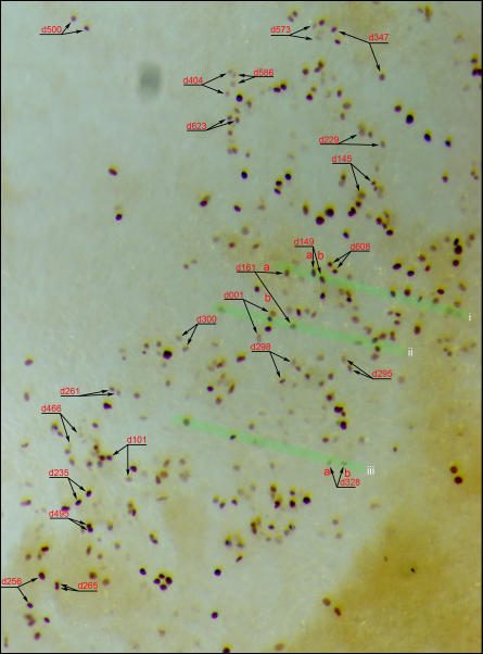
Figure 5. Tracking of labeled cells.
Labeled cells (dividing and post-division) were tracked throughout the time-lapse imaging process and embryos were processed for anti-GFP staining immediately after the last frame. Successfully tracked and matched daughter pairs are shown in this example (whole-mount, anti-GFP stained embryo). The area shown is located in the right-lateral and posterior region of an HH10 embryo. Each daughter pair is also marked with the time of observed mitosis (e.g., d161 represents the division observed at the 161th minute of filming). Three green highlighted stripes (i, ii and iii) indicate regions of sections shown in Fig. 6A and B.

