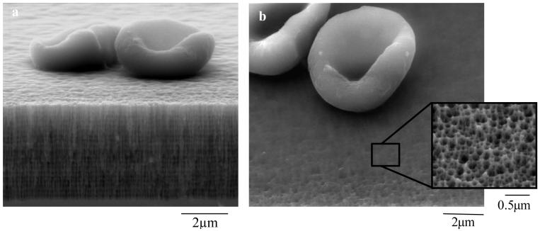
Figure 1. Demonstration of the size -exclusion capability of the porous sensor.
a-b; SEM images of PSi microcavity structure (biosensor substrate) showing erythrocytes filtered out of porous matrix and crosslinked to the surface via glutaraldehyde fixation. a, cross-sectional view. Note the high and low porosity layers in microcavity structure; b, top view with inset showing magnified pores (∼88 nm pore diameter).

