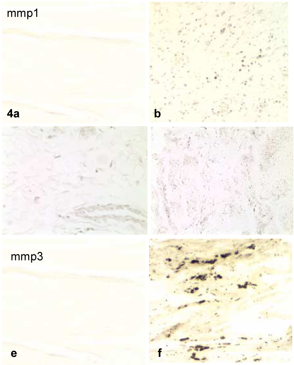
Figure 4.

Examples of control (a,c,e) and contracture (b,d,f) specimens displaying more than 30% of the cells and/or matrix within all sections for MMP-1 (a,b), MMP-2 (c,d), and MMP-3 (e,f). The contracture specimens displayed distinctively more staining than the controls for each cytokine.
