
Figure 5.
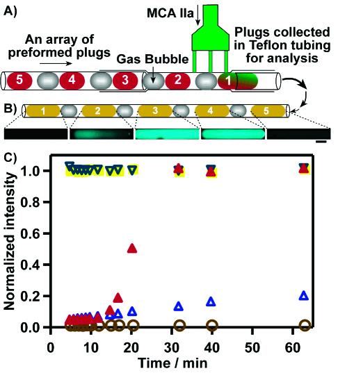
A control blood assay performed with the multijunction device. (A) Schematic drawing of the experimental setup. Substrate MCA IIa was injected into five preformed plugs separated by air bubbles. The five plugs were formed in the order (1, open brown circles) Tris buffer, (2, solid red triangles) human PP, (3, solid yellow squares) thrombin, (4, open inverted triangles) PP + alexin, and (5, open blue triangles) Tris buffer. (B) Fluorescent images of the five resulting plugs taken at the time point of 20 min, scale bar 200 μm. (C) Profiles of fluorescence intensity for five plugs in the assay. Fluorescent images were collected under the same conditions, and the fluorescence intensity was normalized the same way in all images.